Nuclear Medicine
Skip
During nuclear medicine tests, a small amount of radioactive material, called a radiotracer, is used to identify areas in your body that have higher levels of chemical activity. This activity shows up as a bright spot, or “hot spot,” on the nuclear scan and can indicate disease in an organ or tissue. Nuclear medicine can help find problems in their earliest stages.
Nuclear medicine tests are performed on many organs and tissues. Some parts of the process differ with each location, but all tests follow the same basic steps:
- You’re given the radiotracer by mouth, injection or inhalation, depending on the test. The radiotracer requires time to accumulate, which can take anywhere from a few hours to several days, depending on the area being examined. In these cases, you’ll return to the hospital at an appointed time.
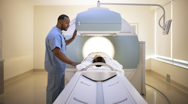
- After the radiotracer accumulates, you’ll lie on an exam table that slides into a circular, donut-shaped device. Special cameras, placed above and below the table, detect the radioactive emissions from the radiotracer.
- A computer uses this information to create images of the area. A radiologist examines the test results and sends a report to your doctor.
Most people who have nuclear medicine tests go home the same day. Doctors often use these tests to find cancer, determine if it has spread, and plan cancer treatments. Nuclear medicine is also used to examine many body areas, including the:
- Heart – To identify abnormal blood flow, measure heart function, determine the extent of damage after a heart attack and evaluate other heart conditions.
- Brain – To investigate problems such as seizures, abnormal blood flow, brain tumor recurrence and Parkinson’s disease.
- Bones – To evaluate fractures, tumors, arthritis, infections and other problems.
- Lungs – To look for blood-flow problems, assess lung function and monitor lung transplants.
- Abdomen – To check for bleeding in the bowel, and abnormalities of the gallbladder, stomach and other abdominal organs.
- Thyroid – To measure function, determine an overactive or underactive thyroid or other condition, and treat thyroid cancer.
Nuclear medicine images are sometimes combined with CT scans in a process called SPECT-CT. This produces a fusion of the two images that can provide more precise information than either test alone.
American College of Radiology – Gold Standard of Accreditation
To achieve the ACR Gold Standard of Accreditation, our facility's personnel qualifications, equipment requirements, quality assurance, and quality control procedures have gone through a rigorous review process and have met specific qualifications. Every aspect of the ACR accreditation process is overseen by board-certified, expert radiologists and medical physicists in advanced diagnostic imaging.